Alnylam Pharmaceuticals is responsible for the funding and content of this website. The site is intended for Healthcare Professionals in Europe, Middle East and Africa. For disease awareness purposes only.
Alnylam Pharmaceuticals is responsible for the funding and content of this website. The site is intended for Healthcare Professionals in Europe, Middle East and Africa. For disease awareness purposes only.
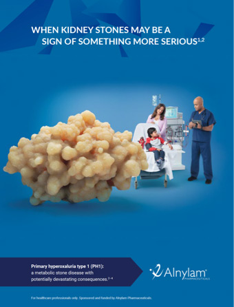
Primary hyperoxaluria (PH) is a group of autosomal recessive metabolic stone diseases resulting from defects in different enzymes involved in glyoxylate metabolism that lead to an overproduction of oxalate in the liver.2,3 There are 3 types of PH: PH1, PH2, and PH3, each of which is caused by a defect in a different enzyme.3
PH1 often presents with kidney stones and is caused by autosomal recessive mutations in the AGXT gene, which impairs the function of the liver-specific enzyme alanine:glyoxylate aminotransferase (AGT).1,3 Normally, AGT processes glyoxylate, which is generated by another liver enzyme, glycolate oxidase (GO).2,3
Do you consider consanguinity when assessing kidney stone patients? Find out why it matters when assessing patients with unexplained kidney stones.
Dr Moochhala: PH1 is an abbreviation that stands for primary hyperoxaluria type 1. So, the primary means that it’s a genetic condition. Hyperoxaluria means there’s an excess of oxalate in the urine. Oxalate is a waste product. And type 1, is telling us that it’s a particular enzyme that has a defect. There’s a number of harmful effects of overproduction of oxalate in the body. The oxalate is deposited in the kidneys, first of all, giving rise to stones, but also giving rise to calcification of the actual kidneys.
It can also be deposited in any soft tissue. So that could be the skin, the eyes, the heart, the muscles, any soft tissue. And that deposition is permanent, so it can reduce the function of that particular tissue. And that’s why it’s such a dangerous, progressive condition that we would like to treat as early as possible. Quite often, the only sign might be that the patient’s having kidney stones over a period of time. It’s a very rare disease. And that’s why it’s so difficult to diagnose.
But we do use clues such as family history. We do see some initial patterns emerging. One of which is we see more a higher prevalence of PH1 in patients from certain backgrounds. And that can include Southeast Asian, Middle Eastern and North African. My message to clinicians anywhere who would see patients to diagnose rare diseases is to think if they’ve got kidney stones, “What caused the stone?” I think in the same way as we would do for any other condition or presentation, we would ask, “Well, what’s the reason?” We should do the same for kidney stone formers. We should ask, “You’re having multiple kidney stones over a period of time?”
“Why is that?” “Have we thought about why that might be?” “Have we investigated it?” “Have we sent you to somebody who might be able to look at that in more detail?” Because that’s the way in which they’re going to make diagnoses of not just common diseases, but also rare diseases. If you think you’re seeing something unusual, such as a patient who’s got maybe a little bit of kidney impairment, but there’s a very strong family history of something going on, or there’s an unusual amount of stone formation in a young patient.
Refer that patient to a nephrologist. Early diagnosis, for PH1, is absolutely essential. The sooner you can start to stabilise the patient’s kidney function and hopefully reduce and even prevent further kidney stone formation. Be persistent. If you think there’s a problem, I would much rather see that patient and find that it was all fine than miss somebody who might potentially have a rare disease, such as PH1 or other rare kidney stone-forming diseases. So my message is to be persistent.
70%–80% of PH cases are PH1. Even so, PH1 is a rare disease, affecting an estimated 1 to 3 cases per million population, an incidence rate of 1 case per 120,000 live births per year in Europe, with higher prevalence in the Middle East and North Africa region.1,3,7
One of the most devastating aspects of PH1 is that it causes a progressive decline in kidney function, culminating in end-stage kidney disease (ESKD).1,3,6 Moreover, there is the potential risk of systemic oxalosis.3,8
Management approaches may lessen damage by reducing stone formation and kidney deposition of calcium oxalate crystals; this underscores the importance of early diagnosis and intervention.3,7,8
PH1 leads to progressive decline in kidney function.2
Liver oxalate overproduction may lead to inflammation and progressive kidney function decline following calcium oxalate crystal formation.8,9 Continuous oxalate overproduction may lead to irreversible damage in the kidneys and other organs.8,9
PH1 advances at variable rates, progressing to ESKD.2 When the kidneys are unable to excrete oxalate effectively due to toxicity from calcium oxalate crystal deposition, systemic oxalosis—the widespread tissue deposition of calcium oxalate—can occur.9 Complications from systemic oxalosis can be fatal.2
In some instances, kidney function can decline after a single incident of dehydration due to acute illness or intense physical activity.10–13 This can occur even in patients with previously stable disease.9,11
PH1 is a genetic disease caused by mutations in the AGXT gene that renders the liver enzyme AGT dysfunctional.3
Normally, AGT processes glyoxylate, which is generated by another liver enzyme, GO.2,3 In PH1, a defect in AGT means glyoxylate is instead converted to oxalate.2 Oxalate cannot be metabolised and is typically excreted by the kidneys at normal levels.3 When overproduced as it is in PH1, oxalate can wreak progressive, irreversible damage.2 Oxalate combines with calcium, creating calcium oxalate crystals.2 These crystals attach to kidney tissues, where they can aggregate to form kidney stones or lead to nephrocalcinosis.2,9 As calcium oxalate accumulates, kidney excretion is impaired, and crystals deposit throughout the body.9

“Given the nature of this disease, PH1 can go undiagnosed for many years. Early symptoms in adults, such as kidney stones, are often attributed to other, much more common conditions. We need to help physicians recognise symptoms faster, to stop progression before a patient reaches end-stage renal failure.”
Kim Hollander
See how genetic testing plays an important role in a PH1 diagnosis.2,4
References: 1. Hoppe B. Nat Rev Nephrol. 2012;8(8):467–475. 2. Milliner DS, Harris PC, Sas DJ, et al. Primary hyperoxaluria type 1. GeneReviews® [Internet]. Updated 15 August 2024. Accessed January 2026. https://www.ncbi.nlm.nih.gov/books/NBK1283/ 3. Cochat P, Rumsby G. N Engl J Med. 2013;369(7):649–658. 4. Cochat P, Hulton SA, Acquaviva C, et al. Nephrol Dial Transplant. 2012;27(5):1729–1736. 5. Ben-Shalom E, Frishberg Y. Pediatr Nephrol. 2015;30(10):1781–1791. 6. Raju DL, Cantarovich M, Brisson ML, et al. Am J Kidney Dis. 2008;51(1):e1–e5. 7. Bhasin B, Ürekli HM, Atta MG. World J Nephrol. 2015;4(2):235–244. 8. Hoppe B, Beck BB, Milliner DS. Kidney Int. 2009;75(12):1264–1271. 9. Leumann E, Hoppe B. J Am Soc Nephrol. 2001;12(9):1986–1993. 10. El-Reshaid K, Al-Bader D, Madda JP. Saudi J Kidney Dis Transpl. 2016;27(3):606–609. 11. Cochat P. Kidney Int. 1999;55(6):2533–2547. 12. Harambat J, Fargue S, Bacchetta J, et al. Int J Nephrol. 2011;2011:864580. 13. Tintillier M, Pochet JM, Cosyns JP, et al. Clin Nephrol. 2004;62(2):155–157.
PH1-INTR-00029 | January 2026
Alnylam Pharmaceuticals is responsible for the funding and content of this website. The site is intended for Healthcare Professionals in Europe, Middle East and Africa. For disease awareness purposes only.
By accessing the website, you confirm to be a Healthcare Professional from Europe, Middle East, or Africa:
If you are not a Healthcare Professional, please access the LivingwithPH1.eu website here.